pPAmCherry-Mito Vector
The mCherry monoclonal antibody detects PAmCherry in the lysate of mammalian cells
PAmCherry is easy to use: Activate PAmCherry in a specific cell or region of a cell, and then track your labeled and activated cells, organelles, or proteins of interest against a dark background
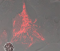
Photoactivated PAmCherry-Mito shows strong red fluorescence and localizes correctly to the mitochondria
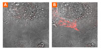
Prior to photoactivation, no red fluorescence is detected in a cell expressing PAmCherry-Mito (Panel A)
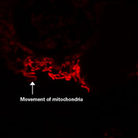
PAmCherry-Mito makes it easy to follow the behavior of a subset of mitochondria