Gene expression quantification for cancer biomarkers

The primary method of diagnosing cancer and disease relies on lengthy, expensive, and complicated histological techniques, which are often employed in patients who are already symptomatic and thus further along in the disease state. Ideally, diagnosis methodologies should be rapid and efficient—enabling affordable and precise screening in presymptomatic, as well as symptomatic, populations. Nucleic acids, either from circulating fluids or FFPE tissue, have emerged as one potential category of biomarkers for studying cancer and disease. Clinically, nucleic acids have already been utilized for prenatal diagnostic testing, and numerous candidate tests are emerging for a variety of disease states. These biomarkers typically fall into four main categories: DNA, mRNA, microRNA (miRNA), and long noncoding RNA (lncRNA).
mRNA markers for many types of cancer, including liver, breast, and prostate have all been reported. However, the high degree of variability and turnover in mRNA makes it a challenging candidate for precision diagnostics. miRNA is a small (typically ~20 nt) species of noncoding RNA that is generally thought to regulate gene expression. Unlike coding mRNAs, miRNAs tend to be relatively stable and resistant to fragmentation. Currently, thousands of unique miRNAs have been identified, with implications across hundreds of diseases, including numerous types of cancers. lncRNAs are longer (typically >200 nt) transcripts that do not code for a specific protein. Similar to miRNA, lncRNA is typically thought to be involved in gene regulation. Hundreds of thousands of lncRNAs have been identified in multiple forms of cancer.
PCR, qPCR, and RT-qPCR are molecular techniques that allow for the detection and quantification of genes and gene expression in a variety of sample sources. These techniques allow for rapid, sensitive, and accurate detection of potential cancer biomarkers.
Highlighted products
Whole-genome SNP array with the Titanium DNA Amplification Kit
Cytogenetic microarrays offer a quick and cost-effective method for the detection of mutations in specific genes, including low-level mosaicism, loss of heterozygosity, and copy number change (Eldai et al. 2013). These mutations are known to affect the prognosis and treatment of various cancers such as melanoma, colorectal cancer, and acute lymphoblastic leukemia. Our Titanium DNA Amplification Kit has been used in microarray assays to screen for and discover mutations in various cancers, such as screening for BRAF mutations in solid tumors with the INFINITI BRAF Assay (Weyant et al. 2014) and uncovering IKFZ1 deletions in childhood acute lymphoblastic leukemia (Lopes et al. 2016). Finally, the high yield and consistent quality provided by the Titanium enzyme enables users of the Affymetrix GeneChip Mapping 500K Assay to achieve the very highest standards in SNP genotyping (Figure 1).

Figure 1. Overview of the GeneChip Mapping 500K Assay.
Quantitative detection of gene expression using RT-qPCR and qPCR kits
 RT-qPCR and qPCR allow for sensitive quantification of genes from RNA and gDNA samples from a variety of sources, including circulating tumor cells (Lianidou 2016). We have developed one-step RT-qPCR kits, two-step RT-qPCR kits, probe-based qPCR mixes, and TB Green-based qPCR mixes in a variety of user-friendly formats for rapid, accurate, and sensitive detection of genes of interest for cancer research. Additionally, as starting materials for cancer research may be precious (e.g., FFPE tissue), we developed our Takara PreAmp Master Mix for exceptional, unbiased preamplification of as little as 12.5 pg of DNA.
These tools have been utilized to detect changes in TrkB signaling in frozen and FFPE uterine leiomyosarcoma samples (Makino et al. 2012), identify key miRNAs in 55 different breast cancer FFPE tissues (Chen et al. 2016), implicate MAF1 in primary human hepatocellular carcinoma tumors (Li et al. 2016), quantify downregulation of the SARI tumor suppressor in prostate cancer tissue and cells, accurately and specifically detect altered expression of RSK4 in multiple cell and tissue samples (Jiang et al. 2017), and ensure reproducible expression of 162 breast cancer-related genes in FFPE tissue samples (view tech note).
We have also developed lyophilized, prevalidated primer sets for colorectal cancer, pancreatic cancer, prostate cancer, melanoma, small cell lung cancer, and non-small cell lung cancer.
Sensitive miRNA quantification with Mir-X miRNA qRT-PCR TB Green kits
Being able to accurately and sensitively quantify rare miRNA is critical to understanding and tracking the signaling network in various types of cancers (Hayes et al. 2014). Our Mir-X miRNA qRT-PCR TB Green kits use a single-step, single-tube reaction to produce first-strand cDNA, which is then specifically and quantitatively amplified using a miRNA-specific primer and TB Green Advantage qPCR chemistry (Figure 2). This enables simple and accurate miRNA detection down to 50 copies from numerous sample types including plasma (Kanemaru et al. 2011), exosomes (Lee et al. 2013), and FFPE tissue (Lee et al. 2013).

Figure 2. Mir-X miRNA qRT-PCR TB Green kit workflow.
High-throughput gene expression analysis with the SmartChip Real-Time PCR System
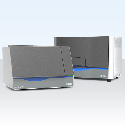
 One of the challenges in screening for these biomarkers is the need to screen hundreds of potential targets in a variety of sample types. High-throughput qPCR has emerged as one potential solution, enabling rapid and sensitive detection of many targets. The SmartChip Real-Time PCR System has been utilized in numerous publications to profile miRNA (Choo et al. 2014), lncRNA (Leucci et al. 2016), and mRNA (Chen et al. 2016) in different forms of cancer and diseases from a multitude of sample types including blood, cell lines, T cells, plasma, and various types of patient biopsies. Critical to all of these studies was the ability of the SmartChip system to support numerous assay and sample configurations, as each disease has a unique and changing set of biomarkers. Through collaboration, we helped researchers design and utilize multiple panels for mRNA, miRNA, and lncRNA that could enable high-throughput screening for key biomarkers in their disease model of interest on the SmartChip system. To learn more about these panels and the research, visit the SmartChip system application pages.
References and product citations
Chen, X. et al. The role of miRNAs in drug resistance and prognosis of breast cancer formalin-fixed paraffin-embedded tissues. Gene 595, 221–226 (2016).
Chen, X. et al. Defining a population of stem-like human prostate cancer cells that can generate and propagate castration-resistant prostate cancer. Clin. Cancer Res. 22, 4505–4516 (2016).
Choo, K. B. et al. MicroRNA-5p and -3p co-expression and cross-targeting in colon cancer cells. J. Biomed. Sci. 21:95 (2014).
Eldai, H. et al. Novel genes associated with colorectal cancer are revealed by high resolution cytogenetic analysis in a patient specific manner. PLoS One 8, e76251 (2013).
Hayes, J., Peruzzi, P. P. & Lawler, S. MicroRNAs in cancer: biomarkers, functions and therapy. Trends Mol. Med. 20, 460–469 (2014).
Jiang, Y. et al. Aberrant expression of RSK4 in breast cancer and its role in the regulation of tumorigenicity. Int. J. Mol. Med. 40, 883–890 (2017).
Kanemaru, H. et al. The circulating microRNA-221 level in patients with malignant melanoma as a new tumor marker. J. Dermatol. Sci. 61, 187–193 (2011).
Lee, J.-K. et al. Exosomes derived from mesenchymal stem cells suppress angiogenesis by down-regulating VEGF expression in breast cancer cells. PLoS One 8, e84256 (2013).
Lee, T. S. et al. Aberrant microRNA expression in endometrial carcinoma using formalin-fixed paraffin-embedded (FFPE) tissues. PLoS One 8, e81421 (2013).
Leucci, E. et al. Melanoma addiction to the long non-coding RNA SAMMSON. Nature 531, 518–522 (2016).
Li, Y. et al. MAF1 suppresses AKT-mTOR signaling and liver cancer through activation of PTEN transcription. Hepatology 63, 1928–42 (2016).
Lianidou, E. S. Gene expression profiling and DNA methylation analyses of CTCs. Mol. Oncol. 10, 431–442 (2016).
Lopes, B. A. et al. COBL is a novel hotspot for IKZF1 deletions in childhood acute lymphoblastic leukemia. Oncotarget 7, 53064–53073 (2016).
Makino, K. et al. Inhibition of uterine sarcoma cell growth through suppression of endogenous tyrosine kinase B signaling. PLoS One 7, e41049 (2012).
Weyant, G. W. et al. BRAF mutation testing in solid tumors: a methodological comparison. J. Mol. Diagn. 16, 481–485 (2014).
Featured products

Unlock answers with nanoscale PCR
Takara Bio's SmartChip ND Real-Time PCR System allows you to flexibly design your own panels, keep costs low, and obtain results in under 3 hr. Sample dispensing and reaction mix distribution is automated for up to 5,184 reactions per chip. Customizable configurations allow 12 to 384 assays to be processed at a time, depending on the number of targets in the panel. The system also simplifies your workflow with full end-to-end software to carry out automated dispensing and qPCR analysis.
Learn moreTakara Bio USA, Inc.
United States/Canada: +1.800.662.2566 • Asia Pacific: +1.650.919.7300 • Europe: +33.(0)1.3904.6880 • Japan: +81.(0)77.565.6999
FOR RESEARCH USE ONLY. NOT FOR USE IN DIAGNOSTIC PROCEDURES. © 2025 Takara Bio Inc. All Rights Reserved. All trademarks are the property of Takara Bio Inc. or its affiliate(s) in the U.S. and/or other countries or their respective owners. Certain trademarks may not be registered in all jurisdictions. Additional product, intellectual property, and restricted use information is available at takarabio.com.


