Using mRNA, miRNA, and lncRNA as biomarkers for cancer and other diseases
The primary method of diagnosing cancer and disease relies on lengthy, expensive, and complicated histological techniques, which are often employed in patients who are already symptomatic and thus further along in the disease state. Ideally, diagnosis methodologies should be non-invasive, rapid, and efficient—enabling affordable and precise screening in presymptomatic, as well as symptomatic, populations. Circulating nucleic acids, sometimes referred to as the circulating transcriptome, have emerged as one potential population of biomarkers for cancer and disease. Clinically, circulating nucleic acids have already been utilized for prenatal diagnostic testing, and numerous candidate tests are emerging for a variety of disease states. These circulating biomarkers typically fall into three main categories: mRNA, microRNA (miRNA), and long noncoding RNA (lncRNA).

Characteristics of different RNA biomarkers
Circulating mRNA markers for liver, breast, and prostate cancers have all been reported. However, extracellular mRNA typically is rapidly degraded to small pieces that are challenging to characterize as unique biomarkers. In some cases, circulating mRNA can form complexes with chaperones, either protein or lipid binding partners, and persist in the blood. However, the high degree of variability and turnover in circulating mRNA makes it a challenging candidate for precision diagnostics.
miRNAs are a small (typically ~20 nt) species of noncoding RNA that is generally thought to regulate gene expression. Unlike coding mRNAs, miRNAs tend to be relatively stable and resistant to fragmentation. Currently, thousands of unique miRNAs have been identified, with implications across hundreds of diseases, ranging from numerous types of cancers, hereditary diseases, heart disease, kidney disease, central nervous system dysfunction and disorders, and obesity, as well as viral and parasitic infections. miRNA has also been found in other body fluids, including saliva, tears, cerebrospinal fluid, and urine.
lncRNA are longer (typically >200 nt) transcripts that do not code for a specific protein. Similar to miRNA, lncRNA is typically thought to be involved in gene regulation. Hundreds of thousands of lncRNAs have been identified and multiple cancers, neurological diseases, heart disease, and aging have been linked to disease-specific lncRNA species.
Solutions for biomarker screening
One of the challenges in screening for these biomarkers is the need to screen hundreds of potential targets in a variety of sample types. High-throughput qPCR has emerged as one potential solution, enabling rapid and sensitive detection of many targets. The SmartChip Real-Time PCR System has been utilized in numerous publications profiling miRNA, lncRNA, and mRNA in various forms of cancer and disease from a multitude of sample types including blood, cell lines, T cells, plasma, and various types of patient biopsies. Critical to all of the studies was the ability of the SmartChip system to support numerous assay and sample configurations, as each disease has a unique and changing set of biomarkers. Through collaboration, we helped researchers design and utilize multiple panels for mRNA, miRNA, and lncRNA that could enable high-throughput screening for key biomarkers in their disease model of interest on the SmartChip system.
High-throughput studies
In some studies, a human oncology panel was utilized. This panel contained over 1,200 gene-specific assays covering 16 functional groups, including signal transduction, cancer, apoptosis, angiogenesis, cardiovascular disease, ADME (toxicology, drug metabolism, and transport), inflammation, kinases, transcription factors, drug targets, G-protein coupled receptors, cell cycle and proliferation, DNA damage repair, growth factors, proteinases, and phosphatases. In one study the human oncology panel was used to study the interaction between human cytomegalovirus (HCMV) and cancer. The study identified 20 differentially expressed genes in multiple pathways including signal transduction (MAPK and PI3K/Akt), inflammation, angiogenesis, tumor suppressor, proapoptotic, and DNA damage repair pathways. These data indicated the potential for HCMV to have oncogenic and/or oncomodulating activity in normally healthy cells. Another study utilized the same panel to study how tyrosyl-tRNA synthetase, a known interactor for protein synthesis, can also localize to the nucleus and regulate DNA damage repair genes.
Other studies utilized an miRNA panel that contained 1,306 pre-validated miRNA targets. The miRNA panel was run on the SmartChip system, which enabled the screening of thousands of miRNA in numerous samples, including colon cancer cells taken from patient tumors and pancreatic ductal adenocarcinoma cells, to identify key miRNA mediators of disease. Outside of cancer, the panel was applied to identify miRNAs linked to Down syndrome in maternal plasma, providing a potential noninvasive method for prenatal diagnosis. The panel has also been utilized to study and characterize different cell populations. One study characterized specific miRNAs shared between cancer cells and stem cells that regulate apoptosis. In another, the subpopulations of miRNA in serum, plasma, and leukocytes were characterized longitudinally. Another group found a mechanism for glucocorticoid suppression of T cells reliant on miRNA-98 by screening treated cells on the SmartChip system.
Finally, some research has studied lncRNAs on the SmartChip system by using a panel containing over 1,700 assays. This panel was developed in collaboration with Biogazelle and designed to be compliant with the Minimum Information for Publication of Quantitative Real-Time PCR Experiments (MIQE) guidelines, as well as curated against genomic databases. In one study, the lncRNA panel was used to study expression in the NCI-60 cancer cell line panel, a set of 60 human cancer cell lines derived from a variety of tissues including brain, blood, bone marrow, breast, colon, kidney, lung, ovary, prostate, and skin. 97% of the screened 1,700 lncRNAs were reproducibly expressed in at least one of the cell lines in the NCI-60 panel. In another study, the lncRNA panel was screened in melanoma samples to identify key mediators of melanomagenesis. This study found that the lncRNA SAMMSON was highly upregulated in the melanoma samples, and led to further study and characterization of its mechanisms. Another recent study utilized the lncRNA panel to develop a screen to look at how somatic copy-number alterations (SCNA) could influence lncRNA alterations to ultimately function as oncogenes or tumor suppressor genes. This panel could prove to be a key method of determining cancer-causing lncRNAs.
Review the citations listed below to see some of the mRNA, miRNA, and lncRNA biomarker research that was enabled by the SmartChip Real-Time PCR System.
Citations
Ammerlaan, W. & Betsou, F. Intraindividual Temporal miRNA Variability in Serum, Plasma, and White Blood Cell Subpopulations. Biopreserv. Biobank. 14:bio.2015.0125 (2016).
Chen, X. et al. Defining a population of stem-like human prostate cancer cells that can generate and propagate castration-resistant prostate cancer. Clin. Cancer Res. 22:4505-4516 (2016).
Choo, K. B., Soon, Y. L., Nguyen, P. N. N., Hiew, M. S. Y. & Huang, C.-J. MicroRNA-5p and -3p co-expression and cross-targeting in colon cancer cells. J. Biomed. Sci. 21:95 (2014).
Davis, T. E., Kis-Toth, K., Szanto, A. & Tsokos, G. C. Glucocorticoids suppress T cell function by up-regulating microRNA-98. Arthritis Rheum. 65:1882-1890 (2013).
Ding, N. et al. BRD4 is a novel therapeutic target for liver fibrosis. Proc. Natl. Acad. Sci. U. S. A. 112:15713-8 (2015).
Kamhieh-Milz, J. et al. Differentially expressed microRNAs in maternal plasma for the noninvasive prenatal diagnosis of down syndrome (Trisomy 21). Biomed Res. Int. 2014 (2014).
Leucci, E. et al. Melanoma addiction to the long non-coding RNA SAMMSON. Nature 531:518-522 (2016).
Mestdagh, P. et al. Evaluation of quantitative miRNA expression platforms in the microRNA quality control (miRQC) study. Nat. Methods 11:809-815 (2014).
Mestdagh, P., Lefever, S., Volders, P. & Derveaux, S. Long non-coding RNA expression pro fi ling in the NCI 60 cancer cell line panel using. Sci. Data 1-6 doi:10.1038/sdata.2016.52 (2016).
Nault, R., Fader, K. A. & Zacharewski, T. RNA-Seq versus oligonucleotide array assessment of dose-dependent TCDD-elicited hepatic gene expression in mice. BMC Genomics 16:373 (2015).
Nguyen, P. N. N., Huang, C.-J., Sugii, S., Cheong, S. K. & Choo, K. B. Selective activation of miRNAs of the primate-specific chromosome 19 miRNA cluster (C19MC) in cancer and stem cells and possible contribution to regulation of apoptosis. J. Biomed. Sci. 24:20 (2017).
Rothwell, D. G. et al. Evaluation and validation of a robust single cell RNA-amplification protocol through transcriptional profiling of enriched lung cancer initiating cells. BMC Genomics 15:1129 (2014).
Sardi, S. H., Glassner, B. J., Chang, J. R. & Yim, S. H. Identification of Critical Biomarkers Responsive to Anti-Autophagy Therapies for Pancreatic Ductal Adenocarcinoma through a Performance Analysis of miRNA Platforms. J. Bioanal. Biomed. 1:doi:10.4172/1948-593X.S10-001 (2014).
Snyder, C. et al. Prevalence of PALB2 mutations in the Creighton University Breast Cancer Family Registry. Breast Cancer Res. Treat. 150:637-641 (2015).
Ullmann, P. et al. Hypoxia-responsive miR-210 promotes self-renewal capacity of colon tumor-initiating cells by repressing ISCU and by inducing lactate production. Oncotarget. 7 (2016).
Volders, P.-J. et al. Targeted genomic screen reveals focal long non-coding RNA copy number alterations in cancer. bioRxiv 113316:doi:10.1101/113316 (2017).
Wei, N. et al. Oxidative stress diverts trna synthetase to nucleus for protection against dna damage. Mol. Cell 56:323-332 (2014).
Wu, L. et al. Full-length single-cell RNA-seq applied to a viral human cancer: applications to HPV expression and splicing analysis in HeLa S3 cells. Gigascience 4:51 (2015).

Unlock answers with nanoscale PCR
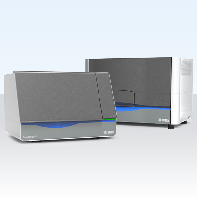
Takara Bio's SmartChip ND Real-Time PCR System allows you to flexibly design your own panels, keep costs low, and obtain results in under 3 hr. Sample dispensing and reaction mix distribution is automated for up to 5,184 reactions per chip. Customizable configurations allow 12 to 384 assays to be processed at a time, depending on the number of targets in the panel. The system also simplifies your workflow with full end-to-end software to carry out automated dispensing and qPCR analysis.
Learn moreTakara Bio USA, Inc.
United States/Canada: +1.800.662.2566 • Asia Pacific: +1.650.919.7300 • Europe: +33.(0)1.3904.6880 • Japan: +81.(0)77.565.6999
FOR RESEARCH USE ONLY. NOT FOR USE IN DIAGNOSTIC PROCEDURES. © 2025 Takara Bio Inc. All Rights Reserved. All trademarks are the property of Takara Bio Inc. or its affiliate(s) in the U.S. and/or other countries or their respective owners. Certain trademarks may not be registered in all jurisdictions. Additional product, intellectual property, and restricted use information is available at takarabio.com.


